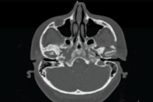
Comparison of brain CT angiography/venography and temporal bone HRCT scan findings in patients with subjective pulsatile tinnitus in affected side and unaffected side
DOI:
https://doi.org/10.15419/bmrat.v5i7.454Keywords:
CT Angiography/Venography, Pulsatile tinnitus, Temporal bone HRCT scanAbstract
Introduction: Pulsatile tinnitus (PT) is a discrete repetitive sound that is synchronous with the patient’s pulse. Incidence of abnormal, often treatable and structural findings in patients with PT has been noted to be high, ranging from 44% to 91%. So, accurate diagnosis of pathologic causes is important for treatment and prognosis.
Objective: The main objective of our study was evaluation of abnormal findings in temporal bone HRCT scan and CTA/CTV in patients affected by pulsatile tinnitus, and comparing these findings with unaffected ear of the same individuals.
Methods: A case-control study was conducted and all the patients who had subjective pulsatile tinnitus with normal physical examinations among outpatient individuals referred to our institution; Rasoul Akram Hospital from September 2015 to august 2017 was included in this study. All patients underwent brain CT Angiography/Venography (CTA/V) and temporal bone high resolution CT scan (HRCT), and their findings were compared.
Results: Thirty patients consist of 20 females and 10 males with mean age of 50.83 years were evaluated in this study. Pulsatile tinnitus was unilateral in all patients except one. Comparing results of CTA/V and temporal HRCT scan in the affected ear showed that temporal HRCT scan in 19(63.3%) out of 30 patients reported normal, while CTA/V only in eight (26.6%) of these patients reported normally. So, capability of CTA/V is statistically significant in diagnosis of abnormalities in the affected ear (P=0.012).
Conclusion: Our findings demonstrate that CTA/V can be a more accurate imaging tool than temporal bone HRCT in initial assessment of patients with subjective pulsatile tinnitus and normal physical examination, and should be done as the first imaging tool in these patients.

Downloads
Published
Issue
Section
License
Copyright The Author(s) 2017. This article is published with open access by BioMedPress. This article is distributed under the terms of the Creative Commons Attribution License (CC-BY 4.0) which permits any use, distribution, and reproduction in any medium, provided the original author(s) and the source are credited.
