Development and evaluation of 12-lead electrocardiogram in the left free wall of accessory pathway localization in patients with typical Wolff-Parkinson-White syndrome
DOI:
https://doi.org/10.15419/bmrat.v5i11.502Keywords:
12-lead ECG, Accessory pathway localization, Left free wall, WPW syndromeAbstract
Objectives: This study was designed to characterize 12-lead electrocardiogram (ECG) for localization of the left free wall lateral accessory pathway (AP) in patients with typical Wolff-Parkinson-White (WPW) syndrome, to develop a new algorithm ECG for localizing APs, and to test the accuracy of the algorithm prospectively.
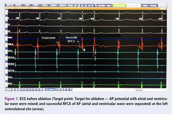
Method: We studied 129 patients; 84 patients had typical WPW syndrome with single anterograde AP identified by successful radiofrequency catheter ablation (RFCA), and were enrolled to build a new ECG algorithm for localizing left free wall APs. Then, the algorithm was tested prospectively in 45 patients and compared with the location of APs successfully ablated by RFCA.
Results: We found that the 12-lead ECG parameters in typical WPW syndrome, such as delta wave polarity in V1, R/S ratio in V1, transition of the QRS complex, and delta wave polarity in inferior, lead to diagnosis and localization of APs, with highest accuracy predicted from 74.5%-100%, and for development of a new ECG algorithm. From the 45 patients who were prospectively evaluated by the newly derived algorithm for the left free wall pathways, the sensitivity and specificity was high (from 75-100%).
Conclusion: The 12-lead ECG parameters in typical WPW syndrome are closely related to left free wall AP localization and can be used to develop a new ECG algorithm by the parameters above. Moreover, the new ECG algorithm can predict the location of APs with high accuracy.

Downloads
Published
Issue
Section
License
Copyright The Author(s) 2017. This article is published with open access by BioMedPress. This article is distributed under the terms of the Creative Commons Attribution License (CC-BY 4.0) which permits any use, distribution, and reproduction in any medium, provided the original author(s) and the source are credited.
