Association of Immunohistochemistry Leptin Expression with Plasma ELISA Leptin Levels in Invasive Breast Ductal Carcinoma: A Cross-Sectional Study
DOI:
https://doi.org/10.15419/bmrat.v10i3.798Keywords:
Breast Cancer, Leptin, Immunohistochemistry, ELISAAbstract
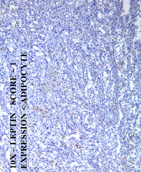
Background: Breast cancer is a leading cause of cancer-related deaths in women globally. Various markers have been identified in breast cancer tissue, including estrogen receptors, progesterone receptors, human epidermal growth factor receptor 2, Ki67, and adipokine. The most crucial mediator in the relationship between obesity and breast cancer is leptin, which encourages the initiation, development, growth, and spread of tumors. Breast cancer development and progression are significantly influenced by leptin, which is present in both blood and tissue. This study aimed to evaluate the association between leptin immunohistochemistry (IHC) expression in tissue sections and leptin plasma levels by enzyme-linked immunosorbent assay (ELISA) in invasive ductal breast carcinomas and correlate this with clinicopathological parameters.
Methods: A laboratory observational cross-sectional study was conducted over 18 months. Tissue sections of invasive ductal breast carcinoma cases were collected for IHC leptin expression, and plasma leptin levels of the same cases were estimated by ELISA. The association of IHC leptin expression and plasma leptin levels with other clinicopathological parameters was determined. All data were entered into a Microsoft Excel spreadsheet and statistical analysis was performed using SPSS 22 software.
Results: Among the study population of 52 cases, 92.3% showed IHC leptin positivity. Plasma leptin levels ranged from 13.21 ? 79.54 ng/ml with a mean of 40.92 ? 20.05 ng/ml. The association of IHC leptin expression and plasma leptin levels was not statistically significant. Among the various clinicopathological parameters studied, the associations of IHC leptin expression with the size (p value-0.04) and stage (p value-0.05) of the tumor were statistically significant. ELISA leptin levels related to parity (p-value 0.04), estrogen receptors (p-value 0.01), and progesterone receptors (pvalue 0.005) were statistically significant.
Conclusion: IHC leptin positivity was identified in 92.3% of cases. The mean plasma ELISA leptin level was 40.92 ? 20.05 ng/ml. IHC leptin expression had a statistically significant association with the size and stage of the tumor, while plasma ELISA leptin levels had a statistically significant association with parity, estrogen, and progesterone receptors.

Published
Issue
Section
License
Copyright The Author(s) 2017. This article is published with open access by BioMedPress. This article is distributed under the terms of the Creative Commons Attribution License (CC-BY 4.0) which permits any use, distribution, and reproduction in any medium, provided the original author(s) and the source are credited.
