A mixture of secretions and extractions derived from antler stem cells heal open wounds in rats with a tendency to leave no scar
DOI:
https://doi.org/10.15419/bmrat.v11i1.855Keywords:
Antler stem cell, Damaged skin, Deer antler, Extraction, Stem cell, Secretions, Mesenchymal stem cellAbstract
Introduction: Deer antlers are remarkable organs as they can regenerate seasonally and leave no scars. Antler-derived stem cell therapy applications are of increasing interest in food, beauty, and medicine.
Method: Antler stem cells (ASCs) were isolated from antlers, and their expression of markers CD73, CD90, CD105, Nanog, and Oct4 was detected by PCR. Their capacity to differentiate into osteoblasts, chondrocytes, and adipocytes was assessed through culture in selection media. In vitro evaluations included wound healing and angiogenic effects using scratch and tube formation assay, respectively. For in vivo assessment, a rat skin defect model was established surgically, and ASC mixtures were applied to the defective skin at a dose of 100μg/100μl. Treatment effects were also evaluated through histological analysis, immunohistochemical staining, and the expression of genes involved in wound healing (including TGF-B1, TGF-B3, TIMP1, Col1, Col3, MMP1, and MMP3) using qRT-PCR method.
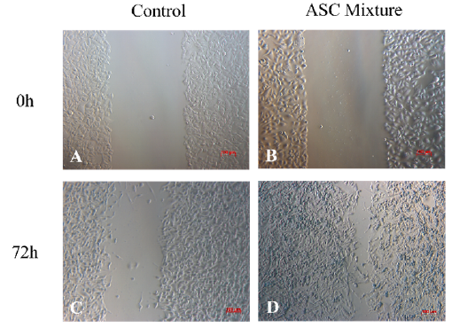
Results: The results indicated that ASCs exhibited fibroblast-like cell shapes and positively expressed specific stem cell markers (CD73, CD90, CD105, Nanog, Oct 4). ASCs demonstrated the capability to differentiate into osteoblasts, chondrocytes, and adipocytes. The products derived from ASCs significantly enhanced NIH-3T3 cell proliferation and stimulated angiogenesis in HUVEC on Matrigel compared to the control group. In the rat skin defect model, wounds treated with the ASC mixture were quicker to heal, starting from day 8, compared to the sham group (day 16). The wound area treated with the ASC mixture was significantly smaller than the control group on day 4 (p < 0.05). Furthermore, the gene expression ratio of TGF-β3/TGF-β1, MMP1/TIMP1, and MMP3/TIMP1 was significantly increased in the ASC group compared to the sham group (p < 0.05).
Conclusion: This study highlights the robust wound-healing efficacy of ASC-derived products.

Published
Issue
Section
License
Copyright The Author(s) 2017. This article is published with open access by BioMedPress. This article is distributed under the terms of the Creative Commons Attribution License (CC-BY 4.0) which permits any use, distribution, and reproduction in any medium, provided the original author(s) and the source are credited.
