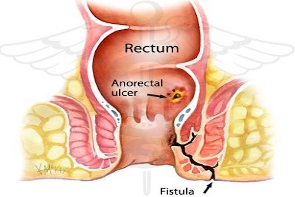
Use of fresh human amniotic membrane for improvement of rabbit perianal surgery wounds
DOI:
https://doi.org/10.15419/bmrat.v4i12.396Keywords:
Amniotic membrane, Fresh human, Perianal wound improvement, Rabbit, MedicineAbstract
Introduction: The use of fresh human amniotic membrane as an accelerating factor for injury repair has been considered for improvement of various injuries, particularly burns, and has shown significant effects. Perianal surgeries, however, have a high recurrence rate due to the anatomy of the area. Thus, finding an effective method in postoperative care may play an important role in improving the quality of surgical procedures in this area. Considering the high recurrent rate of injury in the perianal region and the need for more effective post-surgical care, this study was aimed at evaluating the effects of using amniotic membrane for wound improvement following perianal surgery in a rabbit model.
Methods: Twenty-four New Zealand white male rabbits were divided into two groups. After the rabbits received anesthesia and perianal surgery, group A rabbits received fresh human amniotic membranes (measuring 1x1 cm) to potentially repair the surgical wounds, while group B rabbits did not receive the membranes. The data were analyzed using t-tests and in order to confirm the normal distribution of data, Kolmogorov-Simonov test was used (P=0.03).
Results: In this study, the Abramov surgical wound scoring system was used to determine the wound improvement rate in all specimens. According to the results, this rate was significantly higher in the group administered with amniotic membrane (group A; P=0.0001).
Conclusion: The use of fresh human amniotic membrane plays an important role in the improvement of perianal surgical wounds in rabbits. Thus, this method may be more effective than other methods without membrane anesthetic surgeries.
References
Barlas, M., Gökçora, H., Erekul, S., Dindar, H., & Yücesan, S. (1992). Human amniotic membrane as an intestinal patch for neomucosal growth in the rabbit model. Journal of Pediatric Surgery, 27(5), 597–601. https://doi.org/10.1016/0022-3468(92)90456-H PMID:1625130
Beck, D. E., Roberts, P. L., Saclarides, T. J., Senagore, A. J., Stamos, M. J., & Nasseri, Y. (2011). The ASCRS textbook of colon and rectal surgery. Springer Science & Business Media. https://doi.org/10.1007/978-1-4419-1584-9
Beck, D. E., Wexner, S. D., Hull, T. L., Roberts, P. L., Saclarides, T. J., Senagore, A. J., . . . Steele, S. R. (2014). The ASCRS manual of colon and rectal surgery. Springer Science & Business Media. https://doi.org/10.1007/978-1-4614-8450-9
Drake, R., Vogl, A. W., Mitchell, A. W., Tibbitts, R., & Richardson, P. (2014). Gray’s Atlas of Anatomy E-Book. Elsevier Health Sciences.
Grocott, M., & Pearse, R. (2012). Perioperative medicine: the future of anaesthesia? Oxford University Press.
Haller, G., Laroche, T., & Clergue, F. (2011). Morbidity in anaesthesia: Today and tomorrow. Best Practice & Research. Clinical Anaesthesiology, 25(2), 123–132. https://doi.org/10.1016/j.bpa.2011.02.008 PMID:21550538
Kubo, M., Sonoda, Y., Muramatsu, R., & Usui, M. (2001). Immunogenicity of human amniotic membrane in experimental xenotransplantation. Investigative Ophthalmology & Visual Science, 42(7), 1539–1546. PMID:11381058
Mahajan, M. K., Gupta, V., & Anand, S. (2007). Evalution of fustulectomy and primary skin grafting in low fistula in Ano. JK science, 9, 68–71.
Mohammadi, A.A., and Johari, H.G. (2010). Anchoring sutures: Useful adjuncts for amniotic membrane for skin graft fixation in extensive burns and near the joints. Burns 36, 1134.
Mohammadi, A. A., Sabet, B., Riazi, H., Tavakko-lian, A. R., Mohammadi, M. K., & Iranpak, S. (2015). Human amniotic membrane dressing: An excellent method for outpatient management of burn wounds. Iranian Journal of Medical Sciences, 34, 61–64.
Najibpour, N., Jahantab, M. B., Hosseinzadeh, M., Roshanravan, R., Moslemi, S., Rahimikazerooni, S., . . . Hosseini, S. V. (2013). The effects of human amniotic membrane on healing of colonic anastomosis in dogs. Annals of Colorectal Research, 1(3), 97–100. https://doi.org/10.17795/acr-16139
Phipps, D., Meakin, G. H., Beatty, P. C., Nsoedo, C., & Parker, D. (2008). Human factors in anaesthetic practice: Insights from a task analysis. British Journal of Anaesthesia, 100(3), 333–343. https://doi.org/10.1093/bja/aem392 PMID:18238839
Roshanravan, R., Ghahramani, L., Hosseinzadeh, M., Mohammadipour, M., Moslemi, S., Rezaianzadeh, A., . . . Hosseini, S. V. (2014). A new method to repair recto-vaginal fistula: Use of human amniotic membrane in an animal model. Advanced Biomedical Research, 3(1), 114. https://doi.org/10.4103/2277-9175.131033 PMID:24804188
Schimidt, L. R., Cardoso, E. J., Schimidt, R. R., Back, L. A., Schiazawa, M. B., d’Acampora, A. J., & Sousa, M. V. (2010). The use of amniotic membrane in the repair of duodenal wounds in Wistar rats. Acta Cirurgica Brasileira, 25(1), 18–23. https://doi.org/10.1590/S0102-86502010000100006 PMID:20126882
Smink, D. S. (2015). Schwartz’s Principles of Surgery. Annals of Surgery, 261, 1026. https://doi.org/10.1097/SLA.0000000000001107
Uludag, M., Citgez, B., Ozkaya, O., Yetkin, G., Ozcan, O., Polat, N., & Isgor, A. (2009). Effects of amniotic membrane on the healing of primary colonic anastomoses in the cecal ligation and puncture model of secondary peritonitis in rats. International Journal of Colorectal Disease, 24(5), 559–567. https://doi.org/10.1007/s00384-009-0645-y PMID:19172282
Yetkin, G., Uludag, M., Citgez, B., Karakoc, S., Polat, N., & Kabukcuoglu, F. (2009). Prevention of peritoneal adhesions by intraperitoneal administration of vitamin E and human amniotic membrane. International Journal of Surgery, 7(6), 561–565. https://doi.org/10.1016/j.ijsu.2009.09.007 PMID:19800036

Downloads
Published
Issue
Section
License
Copyright The Author(s) 2017. This article is published with open access by BioMedPress. This article is distributed under the terms of the Creative Commons Attribution License (CC-BY 4.0) which permits any use, distribution, and reproduction in any medium, provided the original author(s) and the source are credited.
