Evaluation effects of ascorbic acid leads to activate and induce osteogenic protein marker expression: in silico and in-vitro study
DOI:
https://doi.org/10.15419/bmrat.v9i1.720Keywords:
ascorbic acid, mesenchymal stem cells (MSCs), prolyl hydroxylaseAbstract
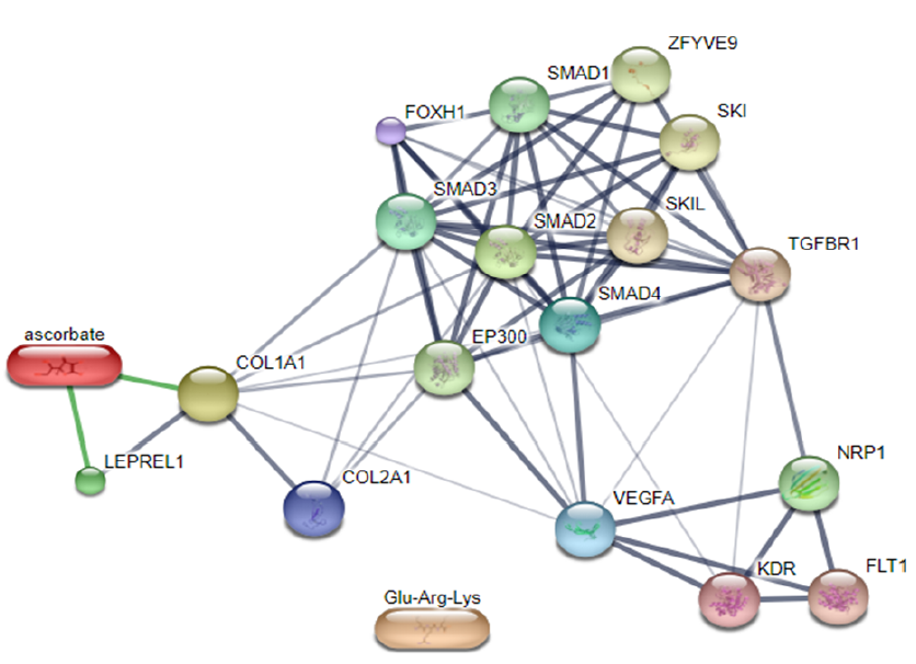
Background: Mesenchymal stem cells (MSCs) are multipotent cells that can differentiate into several cell types including chondrocytes and osteocytes. The process of differentiation into such tissues is aided by transcription factors and collagen gene markers. Collagen biosynthesis is influenced by the presence of vascular endothelial growth factor (VEGF) and ascorbic acid cofactors of the prolyl hydroxylase 2 (PHD2) enzyme found in human cell receptors. The focus of this study is evaluation of how in silico methods can elucidate the effect of ascorbic acid on PHD2 and its expression. This study aimed to evaluate the role of ascorbic acid in its interaction with PHD2 (in silico) and the expression of type 1 collagen (COL1), type 2 collagen (COL2), and VEGF.
Methods: The technique of in silico molecular docking consisted of three steps: preparation of PHD2 receptors using AutoDock tools, preparation of ligand ascorbic acid using AutoDock tools, and molecular docking using AutoDock Vina. The results of in silico molecular docking were visualized using pyMOL and BIOVIA discovery studio. Furthermore, in vitro MSCs from adipose tissue were isolated and characterized based on protein marker expression. The effect of ascorbic acid supplementation (50 and 100 mg/mL) on stem cell differentiation was evaluated based on the mRNA ratio of VEGF/COL1, VEGF/COL2, and COL2/COL1.
Results: Molecular docking of ligand ascorbic acid with PHD2 resulted in nine poses. The third pose was chosen because it had an affinity of -5.7 kcal/mol and a root-mean-square deviation (RMSD) of 1.96, indicating a valid docking result. Meanwhile, the 2D conformer showed van der Waals bonds on Tyr303 and Try329 residues. MSCs were differentiated into adipocytes, chondrocytes, and osteocytes. Regarding the marker ratio of gene expression, at 50 mg/ml ascorbic acid there was an increase in the VEGF/COL1 and VEGF/COL2 ratios and a decrease in the COL2/COL1 ratio compared with the control; at 100 mg/ml ascorbic acid there was a decrease in both ratios (VEGF/COL1 and COL2/COL1), and the VEGF/COL2 ratio continued to increase.
Conclusions: In silico molecular docking between PHD2 and ascorbic acid, which was studied via bioinformatics, resulted in a stable interaction with good ligand position. Further in vitro studies would support the influence of ascorbic acid on transcription factors, collagen gene markers, and the differentiation of MSCs.

Published
Issue
Section
License
Copyright The Author(s) 2017. This article is published with open access by BioMedPress. This article is distributed under the terms of the Creative Commons Attribution License (CC-BY 4.0) which permits any use, distribution, and reproduction in any medium, provided the original author(s) and the source are credited.
