Hyperleukocytosis: a unique cause of an unidentifiable hemoglobin A1c peak
DOI:
https://doi.org/10.15419/bmrat.v11i1.858Keywords:
HbA1c, hyperleukocytosis, T-PLL, assay interferenceAbstract
Background: Glycosylated hemoglobin (HbA1c) serves as a crucial biomarker for the diagnosis and monitoring of diabetes. It can be measured via different methods. Interference during analysis can potentially arise from various factors, including rare occurrences such as hyperleukocytosis.
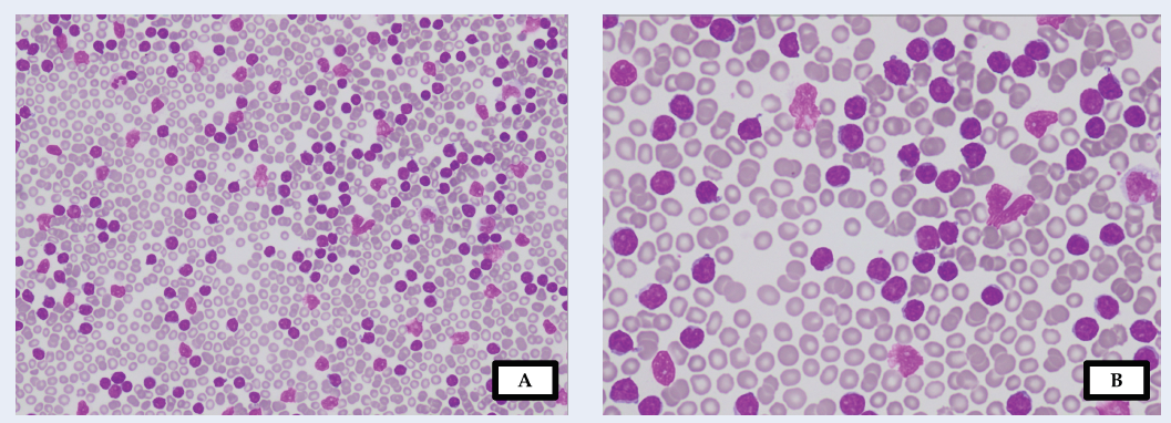
Case presentation: Here, we present the case of a 54-year-old male patient with a 20-year history of type 2 diabetes mellitus who complained of prolonged lethargy, epigastric discomfort, and constitutional symptoms of malignancy. Further investigation revealed a diagnosis of T-cell prolymphocytic leukemia accompanied by hyperleukocytosis, indicated by a white cell count of 574.60 x 10⁹/L with predominant lymphocytes. Chemotherapy and tumor lysis syndrome prophylaxis were initiated. During diabetic monitoring, analysis of HbA1c using capillary electrophoresis revealed an absent HbA1c peak; this has not previously been observed. To address this finding, the sample underwent repeated saline washing and centrifugation. Subsequent analysis demonstrated an improvement, with a well-fractionated HbA1c peak present at 8.7% (71 mmol/mol). Various factors can interfere with HbA1c analysis. Drug and hemoglobin variant interference was ruled out following the recovery of the peak post saline washing. The accelerated migration speed of the sample caused by interfering substances in the plasma was postulated to result in a profile shift, leading to the non-recognition of HbA1c fractions.
Conclusion: By implementing the important step of washing out interfering molecules, the shift was eliminated, allowing for a true HbA1c level measurement. The appearance of an HbA1c peak post saline wash suggests the presence of endogenous substances that interfere with the assay's analytical method.

Published
Issue
Section
License
Copyright The Author(s) 2017. This article is published with open access by BioMedPress. This article is distributed under the terms of the Creative Commons Attribution License (CC-BY 4.0) which permits any use, distribution, and reproduction in any medium, provided the original author(s) and the source are credited.
